Retinal cells
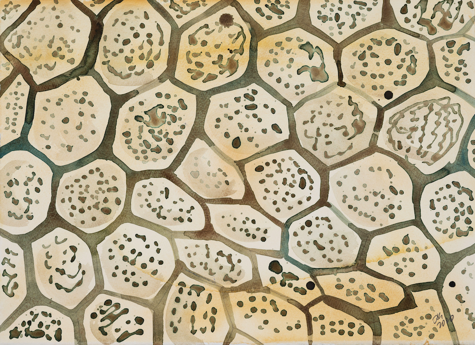
Biorelevant culture of retinal cells on Biolaminin substrates
Effective monolayer differentiation protocol for the generation of hESC-RPE cells under defined and animal origin-free conditions
In a publication by the groups of Drs. Hovatta, Lanner and Kvanta at Karolinska Institutet, Sweden, the authors describe a protocol for effective differentiation of hESC-RPE cells on Biolaminin 521 in a defined and animal origin-free manner (Plaza Reyes, 2015). The differentiated RPE cells exhibit native characteristics including morphology, pigmentation, marker expression, monolayer integrity, polarization, and phagocytic activity. The authors also established a large-eyed geographic atrophy model that allows imaging of the hESC-RPE and host retina. Cells are transplanted in suspension and show long-term integration and form polarized monolayers exhibiting phagocytic and photoreceptor rescue capacity.
In 2020, the same authors published an article in Nature Communications (Plaza Reyes, 2020) where they performed a comprehensive antibody screening and identify cell surface markers for RPE cells. They identified CD140b, CD56, GD2, and CD184 as central cell surface markers to evaluate hPSC-RPE differentiation efficiency, as well as a potential tool for the enrichment of hPSC-RPE during and after differentiation. They show that these markers can be used to isolate RPE cells during in vitro differentiation and to track, quantify and improve differentiation efficiency. Using these markers together with single-cell RNA-sequencing to evaluate the differentiation process, they have established an efficient, robust, direct and scalable xeno-free and defined monolayer differentiation protocol, where culture on supportive human recombinant Biolaminin 111 and 521 eliminates the need for manual selection, allowing large-scale production of pure hPSC-RPE.
The same research group also published an article in Stem Cell Reports (Petrus-Reurer, 2020) where they present a strategy to evade the immune recognition triggered during transplants therapies with human embryonic stem cell-derived retinal pigment epithelial (hESC-RPE) cells. They established single-knockout beta-2 microglobulin (SKO-B2M), class II major histocompatibility complex transactivator (SKO-CIITA) and double-knockout (DKO) hESC lines that were further differentiated into corresponding hESC-RPE lines lacking either surface human leukocyte antigen class I (HLA-I) or HLA-II, or both. The activation of CD4+ and CD8+ T-cells was markedly lower by hESC-RPE DKO cells, while natural killer cell cytotoxic response was not increased. The results show that hESC-RPEs lacking HLA-I and -II evade T-cell response. Moreover, hESC-RPEs lacking HLA-I and -II do not increase NK cell cytotoxic activity. After transplantation of these immune-modified hESC-RPEs in a preclinical rabbit model, donor cell rejection was reduced and delayed. In conclusion, we have developed cell lines that lack both HLA-I and -II antigens, which evoke reduced T-cell responses in vitro together with reduced rejection in a large-eyed animal model.
These results are in accordance with data published by Finnish scientists (Hongisto, 2017) where the authors describe a robust animal origin- and feeder cell-free culture system for undifferentiated hPSCs along with efficient and scalable methods to derive high-quality retinal pigment epithelial (RPE) cells and corneal limbal epithelial stem cells (LESCs). Multiple genetically distinct hPSC lines were adapted to a robust, defined, animal origin-, and feeder-free culture system of Essential 8™ medium and Biolaminin 521 matrix. Thereafter, two-stage differentiation methods toward ocular epithelial cells were established utilizing xeno-free media and a combination of extracellular matrix proteins laminin 521 and Collagen IV. Derivative RPE formed functional epithelial monolayers with mature tight junctions and expression of RPE genes and proteins, as well as phagocytosis and key growth factor secretion capacity after 9 weeks of maturation on inserts. In addition, the authors established xeno-free cryo-banking protocols for pluripotent hPSCs, hPSC-RPE cells, and hPSC-LESCs, and demonstrated successful recovery after thawing on Biolaminin 521 and Collagen IV (Hongisto, 2017).
Biolaminin 521 also supports the culture of human primary RPE cells. Chen et al. show that culturing of human fetal RPE cells on laminin 521 PET transwell inserts creates a model that recapitulates the structural, molecular and apical/basolateral signatures of adult RPE cells (Chen, 2018).
Protocols for ocular cell replacement therapy
The simple animal origin-free methods described by Hongisto et al. could be upgraded to GMP-quality for future preclinical testing and safety and functional efficacy testing of the hPSC-RPE and hPSC LESCs produced with these protocols are currently ongoing in non-human primates and rabbit models of LSCD, respectively (Hongisto, 2017).
Researchers at Cell Cure Neurosciences Ltd. displayed the efficacy of RPE cells derived under defined conditions from clinical and animal origin-free grade human embryonic stem cells following transplantation into the subretinal space of Royal College of Surgeons (RCS) rats (McGill, 2017). Again, the laminin cell culture substrate is being used in the differentiation protocol. The results demonstrate that transplantation of hESC-RPE cells into the subretinal space of RCS rats protected the retinal structure, rescued visual function, preserved rod and cone photoreceptors long-term. OKT was rescued in a dose-dependent manner and the outer nuclear layer (ONL) was significantly thicker in cell-treated eyes than controls. This data combined with data collected in definitive safety studies (tumorigenicity and spiking and safety/biodistribution) has resulted in an FDA approved IND (investigational new drug) and a Phase 1/2a clinical trial for AMD patients is currently ongoing, NCT02286089.
Laminin expression in Bruch’s membrane
It has been shown that the beta-2 chain of the laminin molecule is important for the RPE cells (Libby, 1996). It surrounds RPE cells at the onset of rod photoreceptor birth and is present on the apical surface of the retinal neuroepithelium. At birth, laminin that contains the beta-2 chain fills the matrix between the juxtaposed surfaces of the RPE and neuroepithelium. It is present throughout postnatal development and is also is found in association with blood vessels in the neural retina. A publication by Aisenbrey and colleagues show that the multi-layered extracellular matrix underlying the retina, Bruch’s membrane, contains several laminins that retinal pigment epithelial cells grow on. The main isoforms expressed are laminin 521, 511, 332 and 111. RPE cells adhere robustly to all laminins of Bruch´s membrane and actively synthesize these laminins (Aisenbrey, 2006).
Protocols:
Effective production of hESC-RPE cells in an animal origin-free and defined manner
Production and transplantation of GMP grade animal origin-free hESC-derived RPE cells
Animal origin-free and feeder-free differentiation of human pluripotent stem cells to RPE cells
Succeed with your application
-
Application note 011: RPE differentiation on Biolaminin 521
Clinically compliant retinal pigment epithelial cells from human stem cells
Open pdf -
Xeno-Free and Defined Human Embryonic Stem Cell-Derived Retinal Pigment Epithelial Cells Functionally Integrate in a Large-Eyed Preclinical Model
Plaza Reyes A., Petrus-Reurer S., Antonsson L., Stenfelt S., Bartuma H., Panula S., Mader T., Douagi I., Andre H., Hovatta O., Lanner F., Kvanta A. Stem Cell Reports, 2015
Read more -
Long-Term Efficacy of GMP Grade Xeno-Free hESC-Derived RPE Cells Following Transplantation
McGill T.J., Bohana-Kashtan O., Stoddard J.W., Andrews M.D., Pandit N., Rosenberg-Belmaker L.R., Wiser O., Matzrafi L., Banin E., Reubinoff B., Netzer N., Irving C. Trans Vis Sci Tech., 2017
Read more -
Xeno- and feeder-free differentiation of human pluripotent stem cells to two distinct ocular epithelial cell types using simple modifications of one method
Hongisto H., Ilmarinen T., Vattulainen M., Mikhailova A., Skottman H. Stem cell research and therapy, 2017
Read more -
Instructions 001: Coating with Biolaminin substrates
Protocol and concentration calculations for coating cultureware with Biolaminin
Open pdf
Biolaminin Key Advantages
Biolaminin 521 cultured hESC-RPE cells exhibit native characteristics including morphology, marker expression, monolayer integrity, pigmentation, polarization, and phagocytic activity. Biolaminin 111 and 521 eliminates the need for manual selection, allowing large-scale production of pure hPSC-RPE. Biolaminin culture substrates are a part of FDA approved IND, and a phase 1/2a clinical trial for AMD patients is ongoing.
Specific laminin isoforms are present in different tissue microenvironments and are essential for cell survival, proliferation, and differentiation. Biolaminin products allow you to imitate the natural cell-matrix interactions in vitro.
Our products have consistent composition and quality. This enables minimized variability between experiments and uniform pluripotency gene expression profiles between different cell lines.
All our matrices are chemically defined and animal origin-free, which makes them ideal substrates for each level of the scientific process – from basic research to clinical applications.
Numerous scientists have found our products and finally succeeded in their specific stem cell application. The power of full-length laminins incorporated into various cell systems is well documented in scientific articles and clinical trials.
Recommended products
-
Biolaminin 521 LN (LN521)
Human recombinant laminin 521
Biolaminin 521 LN is the natural laminin for pluripotent stem cells and therefore reliably facilitates self-renewal of human ES and iPS cells in a chemically defined, feeder-free and animal origin-free stem cell culture system. LN521 is animal origin-free to the primary level.View product -

Biolaminin 521 MX (MX521)
Human recombinant laminin 521
Biolaminin 521 MX is the natural laminin for pluripotent stem cells and therefore reliably facilitates self-renewal of human ES and iPS cells in a chemically defined, feeder-free stem cell culture system. MX521 is animal origin-free to the secondary level.View product -

Biolaminin 521 CTG (CT521)
Human recombinant laminin 521
Biolaminin 521 CTG is a full-length, human, recombinant laminin 521 substrate, the only one of its kind on the market, providing an optimal environment for feeder-free culture of human PSCs, MSCs and most anchorage-dependent progenitor cell types. CT521 is animal origin-free to the secondary level and designed for clinical studies.View product -

Biosilk 521
3D culture substrate
Biosilk 521 is a natural biomaterial made of spider silk and laminin 521 – a biocompatible 3D culture mesh for expansion and long-term differentiation of human pluripotent stem cells and for organoid formation of various cell types.View product

Talk to our team for customized support
We are here to help you in your journey.