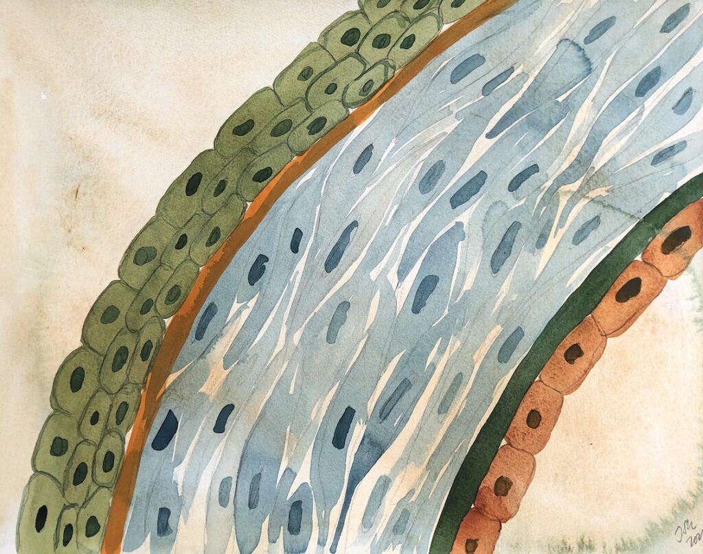
The cornea
Biorelevant culture of corneal cells on Biolaminin substrates
Laminins are an important component of both corneal epithelial and endothelial basement membranes
The cornea consists of three tissue layers: the corneal endothelium, the stromal keratocytes, and the corneal epithelium, separated from one another by a specialized basement membrane. The corneal endothelial cells sit on Descemet’s membrane whereas the corneal epithelial cells sit upon the epithelial basement membrane. Underneath stretches the Bowmans membrane, an acellular, condensed region of the apical stroma, composed primarily of randomly organized yet tightly woven collagen fibrils (Stepp, 2006).
Human corneal endothelial basement membrane contains laminin isoforms 521, 511 and 332
Human corneal endothelial cells (CECs) form a thin cell monolayer of highly polarized flat cells. The basement membrane of the corneal endothelium, also called Descemet’s membrane, is composed of type IV collagen, type VIII collagen, laminin 332, laminin 411, and laminin 511, laminin 521 perlecan and nidogens (Massoudi et al., 2016, Kabosova et al., 2007; Bytröm et al., 2007; Okumura et al., 2015). The Integrin α3β1 receptor is found exclusively at the basal surface of the CECs and ligands of integrin α3β1, such as laminin 332, laminin 511, and laminin 521 are efficient coating substances that improve the yield of in vitroCEC cultures (He et al., 2016, Okumura et al., 2015; Yamaguchi et al., 2011).
In a recent publication, Okumura et al show that Biolaminin 511 and Biolaminin 521 supports the adhesion and proliferation of human CECs. Especially Biolaminin 511 improves the cell adhesion and cell density through the activation of the phosphorylated focal adhesion kinase (FAK). If compared to undercoated control surfaces, human CECs showed a preserved proliferative potential and a higher saturation density when seeded on Biolaminin 511 or Biolaminin 521 (Okumura et al., 2015). In accordance, Hara et al show that corneal endothelial progenitors can be efficiently isolated and expanded on Biolaminin 521 and Biolaminin 511 in a serum-free environment (Hara et al., 2014). The proliferative capacity of these endothelial progenitors is very high on Biolaminin 511 compared to conventional methods.
Laminin in vivo precoating promote the functional recovery of transplanted corneal endothelial cells
Intracameral injection of cultured primary corneal endothelial cells (CECs) are currently being used in clinical trials to treat corneal endothelial dysfunction. However, abnormal adhesion of the grafted CECs affects the application in clinical trials. In a recent study (Zhao, 2020), the authors showed that in vivo coating with Biolaminin 511 (LN511) enhances the adhesion and promotes the functional regeneration of the grafted CECs, improving the therapeutic function for corneal endothelial dysfunction. Rabbit CECs and extracellular matrix of the posterior Descemet’s membrane (DM) were removed by scraping and LN511 were intracamerally injected to form a coating. The injected LN511 could settle and form a coating on the posterior surface of DM and after CEC transplantation, corneal clarity of rabbits in the LN511 group recovered more rapidly compared to the DPBS control group. In addition, the corneal thickness of the LN511 group was significantly lower than the DPBS group. Moreover, LN511 promoted rapid adhesion of the grafted CECs, tight junction formation, and expression of Na+ /K+ – ATPase and ZO-1. In vitro analysis revealed that the functions of LN511 on the cultured human CECs mechanistically depended on the cell density and the nuclear-cytoplasmic translocation of the Yes-associated protein. In summary, LN511 precoating improves the posterior DM, enhances the adhesion of the grafted CECs, and promotes the functional regeneration of CEC transplantation.
The corneal epithelial basement membrane consists of laminin isoforms 332 and 511
The epithelium constitutes the most external layer of the cornea. It is composed of three cell layers: the basal, the intermediate, and the superficial cell layer (Massoudi et al., 2015). The epithelial basement membrane consists of components secreted by the epithelial basal cells, which include type IV collagen, type VII collagen, laminin 332, laminin 511, nidogens, and heparan sulfate proteoglycans (Massoudi et al. 2016; Byström et al. 2007).
The composition of the central cornea basement membrane differs from that of the limbal epithelium. The limbal, but not the central, corneal basement membrane contains laminin 111, laminin 332, and laminin α2β2 chains (Wang 2011). The corneal limbal epithelium niche factors, like laminin 332 can induce transdifferentiation of murine adult hair follicle stem cells into a corneal epithelial-like phenotype (Blazejewska et al., 2009). Moreover, Kabosova et al. reported a compositional difference also between adult and infant corneal basement membranes. Basal epithelial progenitor cells are both found in the central and limbal part of the newborn cornea, but only located in the limbal part of the adult cornea. The infant epithelial basement membrane does not contain laminin α2 and β2 chains, which indicates a developmental regulation of those proteins (Kabosova et al. 2007).
In an article published by Finnish scientists (Hongisto, 2017), the authors describe a robust animal origin- and feeder cell-free culture system for undifferentiated hPSCs along with efficient and scalable methods to derive high-quality retinal pigment epithelial (RPE) cells and corneal limbal epithelial stem cells (LESCs). Multiple genetically distinct hPSC lines were adapted to a robust, defined, animal origin-, and feeder-free culture system of Essential 8™ medium and Biolaminin 521 matrix. Thereafter, two-stage differentiation methods toward ocular epithelial cells were established utilizing animal origin-free media and a combination of extracellular matrix proteins laminin 521 and Collagen IV. In addition, the authors established animal origin-free cryo-banking protocols for pluripotent hPSCs, hPSC-RPE cells, and hPSC-LESCs, and demonstrated successful recovery after thawing on Biolaminin 521 and Collagen IV (Hongisto, 2017).
Laminin expression changes in the corneal basement membranes in eye diseases
Several eye conditions, such as keratoconus, bullous keratopathy, and Fuch’s corneal dystrophy are, are related to the disruption of Bowman’s membrane. In most keratoconus corneas, the Descemet’s membrane (DM) contains laminin 332, normally only found in the epithelial basement membrane. in addition to laminin 511 normal corneas (Bytröm et al. 2007).
In diabetic keratopathy, human corneal epithelial cells show a decrease in laminin α3 chain expression under high glucose conditions, resulting in weakened cell adhesion and (Lu et al. 2006).
Mutations of the laminin α2 chain have been described to be associated with distinct ocular anomalies, including corneal and retinal anomalies, in patients with Pierson syndrome. This demonstrates the importance of the laminin α2 chain for the anterior eye segment development (Zenker et al. 2004).
Protocol:
Succeed with your application
The skin-specific laminin proteins play important roles in the maintenance of keratinocyte phenotypic integrity and promote cell survival.
-
Laminin-511 and -521 Enable Efficient In Vitro Expansion of Human Corneal Endothelial Cells
Okumura N., Kakutani K., Numata R., Nakahara M., Schlötzer-Schrehardt U., Kruse F., Kinoshita S., Koizumi N. IVOS Cornea, 2015
Read more -
Instructions 001: Coating with Biolaminin substrates
Protocol and concentration calculations for coating cultureware with Biolaminin
Open pdf -
Human stem cell based corneal tissue mimicking structures using laser-assisted 3D bioprinting and functional bioinks
Read more
Biolaminin Key Advantages
Laminins are essential components of the corneal epithelial and endothelial microenvironments. Biolaminins 511 and 521 are optimal substrates to facilitate proper adhesion and function of CECs, enabling clinical transplantation.
Specific laminin isoforms are present in different tissue microenvironments and are essential for cell survival, proliferation, and differentiation. Biolaminin products allow you to imitate the natural cell-matrix interactions in vitro.
All our matrices are chemically defined and animal origin-free, which makes them ideal substrates for each level of the scientific process – from basic research to clinical applications.
Our products have consistent composition and quality. This enables minimized variability between experiments and uniform pluripotency gene expression profiles between different cell lines.
Numerous scientists have found our products and finally succeeded in their specific stem cell application. The power of full-length laminins incorporated into various cell systems is well documented in scientific articles and clinical trials.
Recommended products
-
Biolaminin 521 LN (LN521)
Human recombinant laminin 521
Biolaminin 521 LN is the natural laminin for pluripotent stem cells and therefore reliably facilitates self-renewal of human ES and iPS cells in a chemically defined, feeder-free and animal origin-free stem cell culture system. LN521 is animal origin-free to the primary level.View product -
Biolaminin 332 LN (LN332)
Human recombinant laminin 332
Biolaminin 332 supports cells in epithelial basement membranes, lining surfaces of the body such as the skin, hair follicles, oral cavity, gastrointestinal and urinary tract, lungs, and different glands.View product -
Biolaminin 521 MX (MX521)
Human recombinant laminin 521
Biolaminin 521 MX is the natural laminin for pluripotent stem cells and therefore reliably facilitates self-renewal of human ES and iPS cells in a chemically defined, feeder-free stem cell culture system. MX521 is animal origin-free to the secondary level.View product -
Biolaminin 521 CTG (CT521)
Human recombinant laminin 521
Biolaminin 521 CTG is a full-length, human, recombinant laminin 521 substrate, the only one of its kind on the market, providing an optimal environment for feeder-free culture of human PSCs, MSCs and most anchorage-dependent progenitor cell types. CT521 is animal origin-free to the secondary level and designed for clinical studies.View product

Talk to our team for customized support
We are here to help you in your journey.