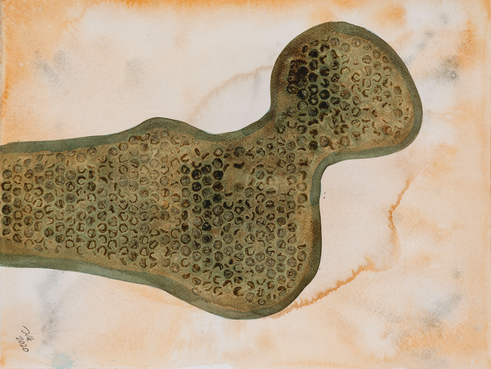
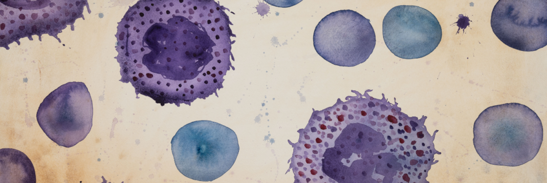
Bone and cartilage cells
Biorelevant culture of bone and cartilage cells on Biolaminin substrates
High expression of laminin isoforms in the bone microenvironment
Bone cells, including include osteocytes, osteoblasts, osteoclasts, osteogenic cells (stem cells), and lining cells, reside in the bone marrow. There are several laminin family proteins expressed in the human bone marrow microenvironment where laminin 411/421 and laminin 511/521 are the most abundant isoforms, synthesized by human bone marrow stromal cells. Laminin isoforms 111, 331, and 332 are also expressed in the human bone marrow microenvironment (Siler, 2000). Laminin 332 is specifically expressed around primary osteoblasts and osteoblast-like cells localized on the bone surface (Uehara, 2017)
LN332 negatively regulates osteoclastogenesis and promote osteogenic differentiation
A publication by German researchers shows that laminin 332 promotes attachment, and compared to plastic, osteogenic differentiation was significantly increased by laminin 332 (Mittag, 2012). In addition, laminin 332 (has been shown to markedly inhibit RANKL induced osteoclastogenesis (Uehara, 2017). The suppression is thought to be mediated through laminin binding to integrin receptors α3β1, α6β1, and α6β4 (Uehara, 2017; Hashimoto, 2006). Laminin 332 is expressed by primary osteoblasts in culture and the laminin γ2 chain is transiently expressed in chondrocytes during development (Uehara, 2017; Hashimoto, 2006). Laminin 332 expression is negatively regulated by osteoclastogenic factors, suggesting that LN332 is a novel negative regulator that may define osteoclastogenesis spatiotemporally in bone tissues (Uehara, 2017). Laminin 332 has also been shown to suppress the chondrogenic differentiation of BM-MSC without inducing apoptosis or inhibiting cell growth (Hashimoto, 2005; Hashimoto, 2006). However, laminin 332 had no effect on the osteogenic differentiation of MSCs (Hashimoto, 2006). These results suggest that laminin 332 may contribute to the development of bone tissues by promoting proliferation and by suppressing the chondrogenic differentiation of MSCs.
LN521, LN511, LN332, and LN111 increase the attachment of bone marrow cells in culture
Laminin 511 and 521 are the most abundant isoforms in the bone marrow. Bone marrow-derived mesenchymal stem cells (BM-MSCs) cultured in vitro have been shown to synthesize α5, α4, α3, α1, and β2 at a significant amount (Seeger, 2015; Siler, 2000; Hashimoto, 2006). Naturally, laminin 511 and 521 have been shown to have strong adhesive interactions with human CD34+ cell lines (Siler, 2000). MSCs does not do not attach well to laminin 111, 211, or 221 (Sun, 2017). Laminin 511, 521, and 332 promote the strongest BM-MSC growth and proliferation rate and affect the mitogenic activity and migration of these cells via binding to integrin α6β1 and α3β1 (Siler, 2000; Sun, 2017, Hashimoto, 2005; Hashimoto, 2006). Yang and Xiao (2016) present a protocol for the culture of bone marrow MSC (BM-MSCs) on laminin 521 and laminin 511. Both laminin isoforms show a significantly faster and stronger attachment compared to uncoated wells and support the seeding of a lower cell number compared to uncoated plats.
When investigating the effects of different ECM coatings for the formation of cell sheets, laminin 521 showed the highest success (Jiang, 2016). Bone marrow mesenchymal stem cells (BMSC) cultured on laminin 521 coated TiO2 nanodot films rapidly attached and spread and formed an intact cell with good viability improved osteogenesis.
Chondrogenic culture of bone marrow-derived mesenchymal stem cells (MSCs) in hydrogels and tissue-engineered cartilaginous constructs reveals extensive expression of laminin throughout the ECM (Sun, 2017). Chondrocytes and rat nucleus pulposus (NP) cells attach strongly to plates coated with laminin isoforms 521, 511, 332, or 111, mainly via the interaction with integrin receptor α6β1 (Sun, 2017). Primary osteoblasts in culture, express laminin 332 which promotes attachment and osteogenic differentiation (Mittag, 2012).
High laminin expression in the cartilage microenvironment
Cartilage is a specialized connective tissue with a multi-component extracellular matrix (ECM) that regulates cartilage repair and regeneration and maintains its functionality. The chondrocytes, the major cartilage resident cells, produce laminins (LN) α1, α2, α3, α4, α5, β1 and γ1 chains, collagen type IV, proteoglycans, elastin, nidogen, and perlecan and these that forms a surrounding thin pericellular matrix (Kvist, 2008; Sun, 2017). There is increasing evidence showing that laminins, secreted by chondrocytes and primarily located in the PCM in cartilage and cartilage-like tissues, are involved in the regulation of chondrocyte activities, such as adhesion, migration, and survival. Furthermore, laminins play a major role in stem cell proliferation and chondrogenic differentiation. Laminins are mainly located in the PCM of human adult articular cartilage with the most prominent staining in the cartilage superficial layer, a niche for chondroprogenitor cells (Kvist, 2008; Sun, 2017; Chen, 2009; Lee, 1997). The influence of the pericellular matrix on the chondroprogenitor cells has been shown to be important for the expression of the major regulator transcription factors, SOX9, and RUNX2 (Schminke, 2016). Similar to the location pattern in the superficial layer of adult articular cartilage, most laminins are detected in the resting zone in epiphyseal cartilage, and expression decreases in the proliferating and hypertrophic zones (Kvist, 2008; Sun, 2017; Kruegel and Miosge, 2010).
Laminin expression changes during human cartilage development
Laminins are continuously expressed during all developing stages of cartilage and cartilage-like tissues and show a region-specific expression pattern. In cartilage from newborn mice, basement membrane components are widespread in the territorial and interterritorial matrix, while in mature cartilage of adult mice the basement membrane components are localized mainly to a narrow pericellular zone around the chondrocytes (Kvist, 2008). Of the laminin subunits surveyed, α1, α5, β1, and γ1 have shown the highest staining intensity (Kvist, 2008). Lee and colleagues found that laminin chains (α1, α2, β1, β2, and γ1), produced by chicken embryo sternal chondrocytes, exhibited an increased expression in aggregated cells during the maturation stage (Lee, 1997). In newborn mouse knee cartilage, laminin 111 showed staining in the extracellular matrix, while laminin α5 (511 and 521) stained weakly at this point (Kvist, 2008). The laminin β1 chain is expressed from gestational weeks (gw) 10 onwards but not during gw 8 and 9, whereas the detection of the laminin β2 chain was limited to gw 8 and 9. This indicates a developmental switch in the laminin β chain and suggests that the laminin β1 chain does not play a role in human cartilage development until the fetal stage. Later at gw 17, a strong pericellular immunohistochemical reaction for LN111 can be detected (Roedinger, 2010). Laminin chains β1 and β2 are also expressed in the cytoplasm of chondrocytes in other species, such as chicken and mice (Roedinger, 2010). Laminin 332 has also been shown to be expressed in embryonic cartilage (Kruegel and Miosge, 2010; Roedinger et al. 2010). The transiently expressed laminin γ2 chain in embryonal cartilage suggests a possible role of laminin 332 in chondrogenesis (Uehara, 2017; Hashimoto, 2006; Lu, 2001).
During the development of intervertebral disc (IVD), laminin is distributed pericellular in developing nucleus pulposus (NP), annulus fibrosus (AF), and vertebral bodies of rats. In general, a gradual shift from diffuse generalized staining of basement membrane components in the newborn towards a pericellular localization in the mature cartilage has been observed (Kvist, 2008). Laminin interactions with nucleus pulposus (NP) cells are distinct from that of the annulus fibrosus (AF) cells. The laminin chain α5 (LN511 or LN521) and corresponding receptors CD239, integrin subunits α3, α6, and β4, are to a higher level expressed in the nucleus pulposus (NP) regions and the α1 chain (LN111 or LN121) shows higher expression in AF cells (Chen, 2009).
Laminin chains are also differentially expressed during chondrocyte differentiation. With the use of an in vitro chick chondrocyte differentiation mode, the expression and localization of fibronectin, laminin, and their receptors were investigated. The results show that fibronectin contributes to the initial cell-cell interactions but is downregulated and replaced by laminin in the in vitro cell condensation process predominantly in the central areas of the cell aggregates, which correspond to differentiating cells. Laminin 111 is maximally expressed at the higher level of cell aggregation and progressively decreases during the differentiation to stage I chondrocytes. The increase in α1, β1, and γ1, LN mRNAs is paralleled by a switch in the isotype of α6 integrin subunits synthesized, from B to A (Tavella, 1997).
The role of laminin in cartilage repair and regeneration
It has been suggested that laminins regulate the fate and functions of chondro-progenitors and chondrocytes in cartilage repair and regeneration. Laminins exist in developing and normal cartilage but many studies show that the expression of laminins significantly decreased or disappears in degenerative, traumatically-damaged cartilage. Interestingly, the diverse expression of laminins in degenerative cartilage indicates their role in cartilage degeneration and suggests that laminin could serve as an early marker for cartilage degeneration. An investigation of cartilage tissue and isolated chondrocytes obtained from patients with late-stage knee osteoarthritis (OA) showed significantly higher expression α1 and α5 laminins compared to healthy cartilage specimens (Schminke, 2016). In addition, chondrogenic progenitor cells (CPCs) in culture produced high levels of laminin chain α1 and α5 and it’s been shown that these laminins promote chondrogenesis and restoration of the chondrocyte phenotype by enhancing collagen type II, COMP, and aggrecan expression, and by down-regulating collagen type II (Schminke, 2016), indicating that laminins have essential roles in promoting chondrogenesis of cartilage-forming cells. In degenerated cartilage sites, the laminin chain α4 has also been shown to be highly expressed and has been suggested to play a deleterious role in cartilage degeneration and might aggravate cartilage damage in osteoarthritis (Sun, 2017). The altered laminin expression pattern and/or decreased in quantity in conjunction with the degeneration of the cartilage-like tissues, implicate the role of laminin in cartilage degeneration.
Succeed with your application
-
Other01/06/2021
Instructions: Laminin protein information
Laminin nomenclature Biolaminin product Name of the protein Old synonyms Chain composition Gene name
-
Bone Marrow Mesenchymal Stem Cells Adhesion Assay
Yang Z. and Xiao R. Bio-protocol, 2016
Read more -
Instructions 001: Coating with Biolaminin substrates
Protocol and concentration calculations for coating cultureware with Biolaminin
Open pdf -
Other30/05/2021
Video: Coating cultureware with Biolaminin substrates
Coating cell cultureware with Biolaminin substrates
Biolaminin Key Advantages
Laminins are secreted by chondrocytes and are involved in the regulation of chondrocyte activities, such as adhesion, migration as well as in survival and chondrogenic differentiation. Chondrocytes and rat nucleus pulposus cells attach strongly to plates coated with laminin isoforms 521, 511, 332 or 111, mainly via the interaction with integrin receptor α6β1. Laminins regulate the fate and functions of chondroprogenitors and chondrocytes in cartilage repair and regeneration.
Laminin 511 and 521 have been shown to have strong adhesive interactions with human CD34+ cell lines and promote bone marrow MSC growth and proliferation. Laminin 332 negatively regulates osteoclastogenesis and promotes attachment and osteogenic differentiation.
Specific laminin isoforms are present in different tissue microenvironments and are essential for cell survival, proliferation, and differentiation. Biolaminin products allow you to imitate the natural cell-matrix interactions in vitro.
All our matrices are chemically defined and animal origin-free, which makes them ideal substrates for each level of the scientific process – from basic research to clinical applications.
Our products have consistent composition and quality. This enables minimized variability between experiments and uniform pluripotency gene expression profiles between different cell lines.
Numerous scientists have found our products and finally succeeded in their specific stem cell application. The power of full-length laminins incorporated into various cell systems is well documented in scientific articles and clinical trials.
Recommended products
-
Biolaminin 521 LN (LN521)
Human recombinant laminin 521
Biolaminin 521 LN is the natural laminin for pluripotent stem cells and therefore reliably facilitates self-renewal of human ES and iPS cells in a chemically defined, feeder-free and animal origin-free stem cell culture system. LN521 is animal origin-free to the primary level.View product -
Biolaminin 511 LN (LN511)
Human recombinant laminin 511
Biolaminin 511 is the natural laminin for mouse embryonic stem cells and allows sustained pluripotency without the need to use feeder cells or differentiation inhibitors like LIF.View product -
Biolaminin 332 LN (LN332)
Human recombinant laminin 332
Biolaminin 332 supports cells in epithelial basement membranes, lining surfaces of the body such as the skin, hair follicles, oral cavity, gastrointestinal and urinary tract, lungs, and different glands.View product -
Biolaminin 111 LN (LN111)
Human recombinant laminin 111
Biolaminin 111 is commonly used as a general attachment protein for many cell types in vitro.View product
Talk to our team for customized support
We are here to help you in your journey.